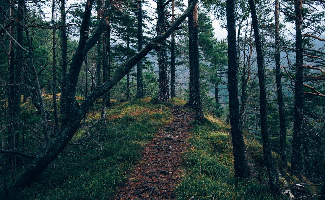
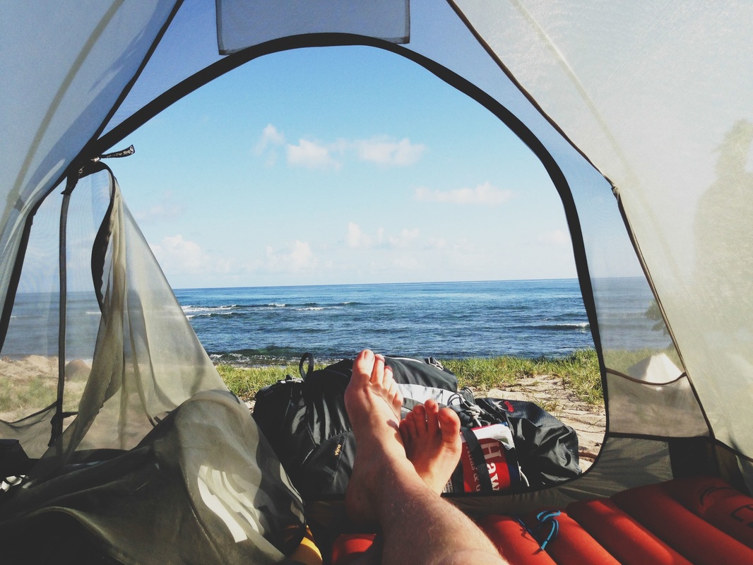
I'm James. This is my year of travel.


Most kidney stones are small enough to be passed in your pee, and it may be possible to treat the symptoms at home with medication. have a medical condition that raises the levels of certain substances in your urineįind out more about the causes of kidney stonesĪfter a kidney stone has formed, your body will try to pass it out when you pee.Over time, the crystals may build up to form a hard stone-like lump. Waste products in the blood can occasionally form crystals that collect inside the kidneys. you have an episode of shivering or shaking.You should contact a GP or NHS 111 immediately if: pain in the side of your tummy (abdomen)įind out more about the symptoms of kidney stones When to get urgent medical help.Larger kidney stones can cause several symptoms, including: You'll usually pee them out without any discomfort. You may not notice if you have small kidney stones. They can be extremely painful, and can lead to kidney infections or the kidney not working properly if left untreated. Kidney stones are usually found in the kidneys or in the ureter, the tube that connects the kidneys to your bladder. They're quite common, with more than 1 in 10 people affected. If left untreated, staghorn calculi result in chronic infection and eventually may progress to xanthogranulomatous pyelonephritis 5.Kidney stones can develop in 1 or both kidneys and most often affect people aged 30 to 60. Staghorn calculi need to be treated surgically, usually PCNL (percutaneous nephrolithotomy) +/- ESWL (extracorporeal shockwave lithotripsy) and the entire stone removed, including small fragments, as otherwise, these residual fragments act as a reservoir for infection and recurrent stone formation. When viewed on bone windows they have a laminated appearance, due to alternating bands of magnesium ammonium phosphate and calcium phosphate 5. Staghorn calculi are radiopaque and conform to the renal pelvis and calyces, which are often to some degree dilated. The collecting system is filled with a densely calcified mass, producing marked posterior acoustic shadowing. The vast majority of staghorn calculi are radiopaque and appear as branching calcific densities overlying the renal outline and may mimic an excretory phase intravenous pyelogram. Uric acid and cystine are the underlying components of a minority of these calculi 5. Struvite accounts for approximately 70% of the composition of these calculi and is usually mixed with calcium phosphate thus rendering them radiopaque on both plain films and CT. 
Urease hydrolyses urea to ammonium with an increase in the urinary pH 3-5. Proteus, Klebsiella, Pseudomonas and Enterobacter). Staghorn calculi are composed of struvite (chemically this is magnesium ammonium phosphate or MAP) and are usually seen in the setting of recurrent urinary tract infection with urease-producing bacteria (e.g. The majority of staghorn calculi are symptomatic, presenting with fever, haematuria, flank pain and potentially septicaemia and abscess formation. Staghorn calculi are the result of recurrent infection and are thus more commonly encountered in women 6, those with renal tract anomalies, reflux, spinal cord injuries, neurogenic bladder or ileal ureteral diversion.
0 Comments
Leave a Reply. |
AuthorWrite something about yourself. No need to be fancy, just an overview. ArchivesCategories |
 RSS Feed
